TMCnet News
OptraSCAN Digital Pathology to Present PD-L1 Pathology AI Tool at AACR 2018 MeetingOptraSCAN On-Demand Digital Pathology® will be presenting their poster "A multi-institutional study to evaluate automated scoring of immunohistochemistry slides for assessment of programmed death-ligand 1 (PD-L1) expression in non-small cell lung cancer" at the upcoming American Association for Cancer Research (AACR) in Chicago, April 14-18, #AACR18. This press release features multimedia. View the full release here: https://www.businesswire.com/news/home/20180412005342/en/ 
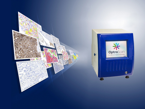
Simultaneously to being scanned, Optra's PD-L1 algorithm quantifies the segmented cells for percent-positive membrane using nuclei as the denominator, resulting in a scoring system for tumor and immune cell percentage, and a processed overlay summary of output images. (Graphic: Business Wire) The study was designed to develop an AI-based Optra automated PD-L1 image analysis system to validate and assess reproducibility for non-small cell lung cancer (NSCLC). In collaboration with Dr. David Rimm, Yale School of Medicine, Dr. Clive Taylor, USC Keck School of Medicine, Dr. Jiaoti Huang, Duke University School of Medicine and Dr. Anagha Jadhav, OptraSCAN. Goal and Methodology The primary goal was to investigate whether PD-L1 immunohistochemistry can be validated and standardized using the OptraSCAN® scanner and scored simultaneously using automated machine-based algorithms. The study assessed interpretation of scores by pathologists, and secondly by Optra's PD-L1 image analysis scoring system to validate reproducibility of automated Optra image analysis for PD-L1 IHC tumor and immune cells. Findings The study findings revealed that Optra PD-L1 whole slide image analysis showed excellent concordance with the pathologists' average scores for tumor (0.83) and immune cell scores (0.6) respectively. These findings suggest promise for the use of automated whole slide image analysis for accurately generating scores necessary for fast, accurate and reproducible results that are geneally cumbersome and time-consuming for pathologists to perform manually. "Previous studies have shown that pathologist-based analysis lacks reproducibility, especially for immune cell expression," says Dr. Anagha Jadhav, Director of Digital Pathology at OptraSCAN. "We designed a study to validate the reproducibility of Optra whole slide PD-L1 image analysis automated machine-based analysis for lung cancer. Our findings demonstrate how computer aided analysis can benefit the accuracy of PD-L1 analysis in a cohort of lung cancer patients, which was otherwise challenging to arrive at an accurate percentage score for both tumor and immune cells within a heterogeneous cell population in the tumor microenvironment." "There is evidence that the efficiency, accuracy and reproducibility of pathologic diagnosis can be enhanced by combining the traditional skills of the pathologist with the use of the 'Intelligent Microscope,' namely a digital scanner with AI software, serving in the role of a pathologist assistant," says Dr. Clive Taylor, Consulting CMO at OptraSCAN. "An immediate impact is (shown) in the scoring of complex Companion Diagnostics, exemplified by PD-L1, where AI algorithms can match the performance of experienced pathologists, as exemplified by this recent OptraSCAN sponsored study." For more information about OptraSCAN's image analysis solutions, visit the company's website here. About OptraSCAN OptraSCAN® (www.optrascan.com) RUO, is the first On-Demand Digital Pathology® System to serve as an affordable tool for transition from conventional microscopy to Digital Pathology, for the effective acquisition of whole slide images, viewing, storing, sharing, consulting, analysis and management of digital slides and associated metadata. OptraSCAN On-Demand Digital Pathology® system includes a small-footprint, low and high throughput WSI (News - Alert) scanner OptraSCAN® (for brightfield, fluorescence and frozen sections/live view mode), an integrated image viewer and image management system ImagePath® and telepathology TELEPath™, CLOUDPath® cloud-based LIMS, image analysis OptraASSAYS™ and CARDS™ (computer aided region detection system), and includes up to 10 TB of complimentary cloud storage. Follow OptraSCAN on Linkedin and Twitter.
View source version on businesswire.com: https://www.businesswire.com/news/home/20180412005342/en/ |

